Colectomy
Colectomy is the most common procedure performed to remove the cancer cells. It is the surgical resection of all or part of the large intestine. It is also called large bowel resection. Colectomy is performed under general anaesthesia by open surgery method or by laparoscopic method. The procedure takes about 1 to 4 hours.
Open colectomy
In this technique a single large incision of about 6 inches is made in the lower abdominal wall. The diseased part of the colon is removed and the healthy ends are sutured. If no healthy large intestine is left, an opening or a small incision called a stoma is created on the abdominal wall. The open end of the large intestine is stitched to the skin of the outer wall of the abdomen. Wastes will pass through this opening into a bag that is attached outside the body. This procedure is called colostomy.
Laparoscopic colectomy
It is a minimally invasive technique where several small incisions are made rather than one large incision. Three to five small incisions are made on the lower abdomen. A laparoscope, a telescopic video camera is used to see the inside of the abdomen, is inserted through an incision. Small surgical instruments are passed through other 2 incisions and colon is removed. Carbon dioxide (CO2) gas is filled in the lower abdomen and expanded for easy access and the diseased part of the colon is removed. The healthy ends are reattached and all the incisions are closed with the sutures.
Complications of colectomy procedure include infection at the site operated, bleeding, and damage to nearby organs.
Some of the ways to prevent the colon cancer include high fibre diets and vitamins, avoid smoking and alcohol consumption, lose weight in case you are obese, and a healthy life style can lessen risk of colon cancer.
Haemorrhoidectomy
Haemorrhoids are masses or lumps formed due to swollen blood vessels inside or outside the rectum. In severe stages, they may become infected or protrude from the anus (prolapsed haemorrhoid) and require surgical removal. The procedure used to treat or remove haemorrhoids is termed Haemorrhoidectomy.
Haemorrhoidectomy is performed under general, regional or local anaesthetic depending on the procedure. The different types are:
- Closed Haemorrhoidectomy: This procedure is used for the removal of internal haemorrhoids. Your doctor uses scissors, scalpels, electro cautery or laser to excise the haemorrhoids and seal the wound with absorbable sutures. It may be associated with complications such as wound infection, bleeding, faecal incontinence (bowel leakage) and anal strictures (narrowing of the anal passage).
- Open Haemorrhoidectomy: This procedure is similar to closed Haemorrhoidectomy, except that the wound is not closed but instead, it is left open. This is done if wound closure is difficult due to the location of the haemorrhoid and if the risk of infection is high.
- Stapled Haemorrhoidectomy: This procedure is used for prolapsed haemorrhoids. Your surgeon uses a circular stapling device to cut off the excessive haemorrhoid tissue and lift the haemorrhoid within the rectum. This stops blood supply to the haemorrhoids so that they shrink and dissolve.
- Rubber band ligation: A rubber band is tied around the base of the haemorrhoid to cut off blood supply so they disappear in a few days. This may be performed in your doctor’s office without the need for anaesthesia.
Pilonidal Sinus
The word pilonidal means a nest of hairs, and a sinus tract is an abnormal narrow channel or cavity in your body. Pilonidal sinuses are infected tracts beneath your skin that predominantly occur at the end of your tailbone, an area called the natal cleft where the buttocks separate.
The exact cause of pilonidal sinus is unknown. It is believed that ingrown hairs combined with changing hormones (pilonidal sinuses occur after puberty), friction (sitting for a long time or from clothing), and infection (ingrown hair and friction can irritate the skin and cause inflammation leading to bacterial invasion) contribute to pilonidal sinus. Having a sedentary job which requires a lot of sitting, obesity, having a deep hairy natal cleft, or a family history of pilonidal sinuses can make you more prone to this condition.
A pilonidal sinus may not have any symptoms at first or can start off as a small painless lump. Once it is infected, it becomes inflamed, fills with pus and develops into a painful cyst. Over a few days you may experience symptoms like:
- Pain, redness, and swelling
- Blood, pus or foul smelling drainage
- Protruding hair from the cyst
- Fever (not seen in everyone)
The symptoms can be rather quick in onset once infection occurs. Pain can be severe and may be experienced whether sitting or standing. Pain can affect your ability to perform your daily activities and can worsen without treatment. Discharge of pus usually releases the pressure and eases the pain, but if the infection is not treated, it can lead to recurrence of the pilonidal sinus.
When you present with symptoms of a pilonidal sinus, your doctor will perform a detailed physical examination. You will be asked for your medical history, which would include information regarding any drainage or change in appearance of the affected area. Your doctor will perform a visual examination to look for tenderness, redness, swelling and inflammation around your affected area. Your doctor may also order a blood test to check for increased white blood cells, which may be an indication of an infection. In rare cases, your doctor will recommend a CT scan to confirm on your condition. Your doctor may conduct tests to rule out other conditions such as a dermoid cyst (tumour of the germ cell) that can resemble a pilonidal cyst.
Treatment may not be necessary if your pilonidal sinus does not become infected. The area can be kept clean and dry, and the surrounding hair removed by shaving or with hair removal creams. However, infected pilonidal sinuses are treated with surgery. Your doctor may perform any of the following procedures:
- Incision and Drainage: This procedure involves cutting open the sinus tract and draining the pus from the abscess. This is a minor procedure and is usually performed under general anaesthesia. You will be able to return home either on the same day of the procedure or the day after. A dressing will be applied, and you will have to visit your doctor’s office daily for dressing changes.
For recurrent infections of your pilonidal sinus, you may be recommended to undergo surgery to remove the sinus and prevent further infections with any of the following procedures:
- Wide Excision: During this procedure, your surgeon will surgically remove a section of your skin along with the infected sinus. This will reduce your chances of a reinfection. Your wound will be left open and dressings will be applied. Your wound may take a long time to heal and you may need to change your dressings daily for two or three months.
- Excision and Primary Closure: During this procedure, your surgeon will surgically remove your affected sinus along with the surrounding skin and then close and seal the wound with stitches. This will help the wound heal quickly; however, the chances of a reinfection are higher when compared to wide excision. A flap of your own skin will be used to close your wound.
Small Bowel Surgery
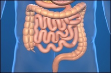
The intestine, a part of the digestive tract, is a long continuous tube, extending from the stomach to the anus consisting of the small intestine, large intestine and rectum.
The small bowel(intestine) which is about 20 feet long and 1 inch in diameter is the largest part of the digestive tract that is divided into three parts – duodenum, jejunum, and ileum. The small intestine folds several times to fit inside the abdomen and plays a vital role in digestion of food and absorption of nutrients and water from the digested food into the body.
There are a number of diseases that can affect the function of the small intestine. Some of these include:
- Bleeding, Infection or ulcers
- Cancer
- Crohn’s disease (inflammatory bowel disease)
- Intestinal blockage or obstruction
- Injury to small intestine
Symptoms
The types of symptoms depend on the condition affecting the small intestine. However, a few common symptoms include nausea, abdominal pain and cramping, rectal bleeding and weight loss.
Diagnosis
Small intestine disorders and other common diseases share some common symptoms making the diagnosis difficult. However, along with a detailed history and physical examination, a few tests may be performed that include CT scan, barium enema X-ray and capsule endoscopy.
Surgical Treatment
While a few conditions of the small intestine like inflammation or infection can be treated with medicines, obstructions or cancer usually require surgery. Small intestine surgery can be done using one of two methods:
- Open technique: Your surgeon will make a 6 inch long incision in the abdomen where the diseased portion of the small intestine is located.
- Laparoscopic technique:Your surgeon will make 3-4 small incisions in the abdomen. One of the incisions may be enlarged (2-3 inches) so as to allow one hand to be placed within the abdomen and remove the diseased segment of the small intestine. The laparoscope (a small, thin tube with a light and tiny video camera that helps visualize the organs during the operation) is inserted into the body through one of the incisions. The television monitor will guide the surgeon to insert other surgical instruments through the other incisions. Air or carbon-dioxide is injected into the abdomen to inflate the abdominal cavity so that the internal organs can be visualized easily.
In both the techniques, the diseased section of the small intestine is clamped off and then removed. The open ends of the small intestine are then connected together using stitches or staples. If there are not enough healthy intestines to re-connect the ends, an ileostomy may be performed. In this procedure, an opening called a stoma is created in the abdominal wall and then the end portion of the intestine close to the stomach is attached to the stoma. The contents of the small intestine are drained out through the stoma into a sealed pouch or bag attached to the abdomen. An ileostomy may be either temporary or permanent.
Risks and Complications
The potential risks and complications of having small intestine surgery include:
- Bleeding in the abdomen
- Frequent diarrhoea
- Injury to the adjacent organs
- Protrusion of the intestine through the incision into the abdomen causing incisional hernia
- Intestinal blockage caused by scar tissue that can develop in the abdomen
- Short bowel syndrome that causes problems with absorption of vitamins and nutrients
- Leakage at the region where the open ends of the intestine are sewn together
- Problems with the stoma, the opening created in the abdominal wall
- Opening of the incision (dehiscence)
- Infection of the incision
Stomach Surgery
Gastric Cancer Surgery
Gastric cancer is the cancer that develops from the cells of the inner layer of the stomach. Cancer is the uncontrolled growth of abnormal cells. The accumulation of these extra cells forms a mass of tissue called tumour. Gastric cancer refers to the cancer of the stomach only and does not include the other organs in the abdomen. Cancer can occur in any part of the stomach. Stomach cancer are of different types depending on the cells of the stomach from which they originate such as hormone making cells, cells of the inner lining or the immunological cells of the stomach. Gastric cancers can spread to the other parts of the body (malignant). They first spread from the stomach to the lymph nodes and then further spread through the lymphatic system. In latter stages, it may spread to other organs such as liver, bones and lungs through blood.
Lymph nodes are the small kidney shaped structures which are the sites for formation of body’s defense cells lymphocytes. Lymph is a clear fluid that flows through lymph vessels and lymph nodes. The interstitial fluid (fluid found between the cells) bathes all the cells and carries excretory products, bacteria and viruses from the cells and helps remove them from the body’s tissues.
Gastric cancer treatment depends on the type and the stage or spread of the cancer. Your age and general state of health is also important consideration by the doctor in determining the treatment modality. Treatment involves surgery, chemotherapy and radiation therapy. Surgery is the most common treatment for gastric cancer.
Surgery
Gastric cancer can spread to other tissues and organs therefore removal of the cancerous tissue is a must. Three kinds of surgeries are used to treat gastric cancer. The type of surgery depends on where the cancer is located and how deep the cancer cells have invaded the area in the stomach. The three types of surgery for gastric cancer are:
- Endoscopic mucosal resection: It is done only when the cancer is detected at an early stage, where the chance of it spreading to lymph node is very less. In this procedure cancerous tissue is removed from the stomach using endoscope, a long flexible tube with the camera at the end. During the procedure an endoscope is passed through the mouth into the stomach and surgical tools are also passed through it to remove the cancerous tissue. The surgery is done using these tools and does not involve any cuts on the body.
- Subtotal gastrectomy: As the name suggests in this surgery a part of the stomach is removed. It is mostly used when the cancer is only in the lower part of the stomach or the upper part of the stomach. During the procedure only part of the stomach is removed, sometimes a part of the oesophagus is also removed along with it. Nearby lymph nodes may also be removed. The remaining part of the stomach is then reattached.
- Total gastrectomy: In this surgery the whole stomach is removed along with nearby lymph nodes. And new stomach is recreated by the small intestine. This is usually done when the cancer has spread to the whole of stomach. Sometimes nearby organs are also removed if the cancer has spread to them.
Chemotherapy and radiation may also be given after the surgery to kill the few cancer cells that may have left after the surgery and to prevent the recurrence of the disease.
Possible complications of surgery for stomach cancer include bleeding, formation of blood clots, and damage to nearby organs. You may also develop frequent heartburn, abdominal pain (especially after eating), and vitamin deficiencies.
After the surgery follow up is very important since the recurrence of the disease is a possibility. In case of any suspicion the doctor will ask for certain tests to confirm his findings. Absorption of vitamin B12 occurs through upper part of the stomach. If upper part of the stomach is removed by surgery Vitamin B12 levels are closely monitored and Vitamin B12 injections are given when required. The doctor may refer you to a nutritionist to plan your diet and you need to eat more often and small meals as the size of the new stomach is small.
Surgery for Acid Reflux
Acid reflux, also called gastro-oesophageal reflux disease (GORD), is a condition where the stomach contents (food or liquid) rise up from the stomach into the oesophagus, a tube that carries food from the mouth to the stomach.
Normally the stomach contents do not enter the oesophagus due to constricted LES. But in patients with acid reflux stomach content travels back into the oesophagus because of a weak or relaxed lower oesophageal sphincter (LES). Lower oesophageal sphincter is a ring of muscle fibers that surrounds the lower-most end of the oesophagus where it joins the stomach. LES acts like a valve between the oesophagus and stomach preventing food from moving backward into the oesophagus
Heartburn is usually the main symptom; a burning-type pain in the lower part of the mid-chest, behind the breast bone. Other symptoms such as a bitter or sour taste in the mouth, trouble in swallowing, nausea, dry cough or wheezing, regurgitation of food (bringing food back up into the mouth), hoarseness or change in voice, and chest pain may be experienced.
The exact cause of what weakens or relaxes the LES in GORD is not known, however certain factors including obesity, smoking, pregnancy, and possibly alcohol may contribute to GERD. Common foods that can worsen reflux symptoms include spicy foods, onions, chocolates, caffeine containing drinks, mint flavorings, tomato based foods and citrus fruits. Certain medications can also worsen the reflux.
There are several tests that can be performed to diagnose acid reflux and they include:
- Endoscopy: This test allows the doctor to examine the inside of the patient’s oesophagus, stomach, and portions of the intestine, with an instrument called an endoscope, a thin flexible lighted tube.
- Barium X-rays: These are diagnostic x-rays in which barium is used to diagnose abnormalities of the digestive tract. You are asked to drink a liquid that contains barium. The barium coats the walls of the oesophagus and stomach and makes the abnormalities visible more clearly. Then X-rays are taken to see if there are strictures, ulcers, hiatal hernias, erosions or other abnormalities.
- Twenty four-hour pH monitoring: In this procedure, a tube will be inserted through the nose into the oesophagus and positioned above the LES. The tip of the tube contains a sensor which can measure the pH of the acid content refluxed into oesophagus. A recorder, strap-like device that can be worn on wrist, will be connected to record the pH of the acid content. The tube will be left in place for 24 hours. Patients can also go back home and perform their regular activities and can record the pH of the acid content when they experience the symptoms. On the next day the recorder will be connected to a computer and the data will be analysed.
- pH Capsule: It is a new method of measuring acid exposure in the oesophagus. A small wireless capsule which is introduced into the oseophagus by a tube through the nose or mouth. The tube is removed after the capsule is attached to the lining of the oesophagus. The pH sensor transmits signals to a computer which collects the data about the acid exposure over the usual 24 hours. The capsule falls off of the oesophagus with time and is passed in the stool.
- Impedance study: This test is similar to pH test but requires two probes; one is placed in the stomach and the other just above the stomach. The dual sensor helps to detect both acidic and alkaline reflux.
Antacids are over-the-counter medicines that provide temporarily relief to heartburn or indigestion by neutralizing acid in the stomach. Other medications such as proton pump inhibitors and H2 antagonists may be prescribed to reduce the production of acid in the stomach.
Surgery may be an option for patients whose symptoms do not go away with the medications. Nissen’s fundoplication is a surgical procedure in which the upper part of the stomach is wrapped around the end of your oesophagus and oesophageal sphincter, where it is sutured into place. This surgery strengthens the sphincter and helps prevent stomach acid and food from flowing back into oesophagus.
Endoluminal gastroplication or endoscopic fundoplication technique requires the use of an endoscope with a sewing device attached to the end, known as EndoCinch device. This instrument place stitches in the stomach below the LES to create a plate which helps reduce the pressure against the LES and help strengthen the muscle.
Chronic GORD left untreated can cause serious complications such as inflammation of the oesophagus, oesophageal ulcer, narrowing of the oesophagus, chronic cough, and reflux of liquid into the lungs (pulmonary aspiration). Some people develop Barrett’s oesophagus, in which there is changes in the oesophageal lining that can lead to oesophageal cancer.
General measures the patient can take to reduce reflux are:
- Avoid eating before going to bed as this may decrease the acid production
- Eat smaller and more frequent meals
- Lose weight if you are over weight
- Elevate the head of the bed
- Eliminate the foods that increases the reflux
- Avoid smoking and use of alcohol
- Check with the physician regarding side effects of prescription medications
Splenectomy
Splenectomy is the surgical removal of spleen, a large organ located in the upper left part of the stomach that contains macrophages, specialized cells that fight against the foreign bodies. Splenectomy is indicated in splenomegaly, a condition of enlarged spleen. Patients with splenomegaly may or may not exhibit the symptoms and will be diagnosed by the physicians by physical examination or radiological diagnosis. The common symptoms include abdominal pain, hiccups, unable to have a large meal, weakness, fatigue, frequent infections, and severe bleeding.
Splenectomy may be performed by open surgery or laparoscopic procedure.
- Open splenectomy: It is a surgical procedure to remove the spleen where the spleen is enlarged and damaged. It is performed under general anesthesia. A large cut is made in the middle or on the left side of the abdomen, below the ribs. The blood vessels are tied. Surgeon removes the spleen and the incisions are stitched after checking for bleeding.
- Laparoscopic splenectomy: It is performed under general anesthesia. It uses a laparoscope, an instrument with a tiny camera and a light at the end. Three to four incisions are made on the abdomen, and the laparoscope is inserted through one of the incisions. The laparoscope allows viewing the area on a bigger screen. Other surgical instruments are inserted through the other incisions. Gas is pumped to expand the abdomen to give more space to work. Spleen is removed using the laparoscope and other instruments. The small incisions are stitched.
Some of the complications include bleeding, wound infection, pneumonia, and injury to other structures.